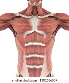
Anatomy Of The Upper Chest Area / Sternum Popping Treatment Pain Chest Pain And Symptoms. The major muscle in the chest is the pectoralis major. Chest a man's chest — like the rest of his body — is covered with skin that has two layers. Anatomy of the chest area. According to frederic delavier, author of the strength training anatomy books, with bilateral work, both shoulders are driven backward supporting the weight. A heart attack results from blocked blood flow, often from a blood clot, to your heart muscle.
A heart attack results from blocked blood flow, often from a blood clot, to your heart muscle. The pectoral region is located on the anterior chest wall. Upper anterior muscles anatomy the science of human anatomy by bartholomeo eustachi, depicting the shape, size. 12 photos of the anatomy of the chest area. Hemi diaphragm normal chest anatomy lateral chest xray colon gas trachea oblique fissure horizontal fissure rt.
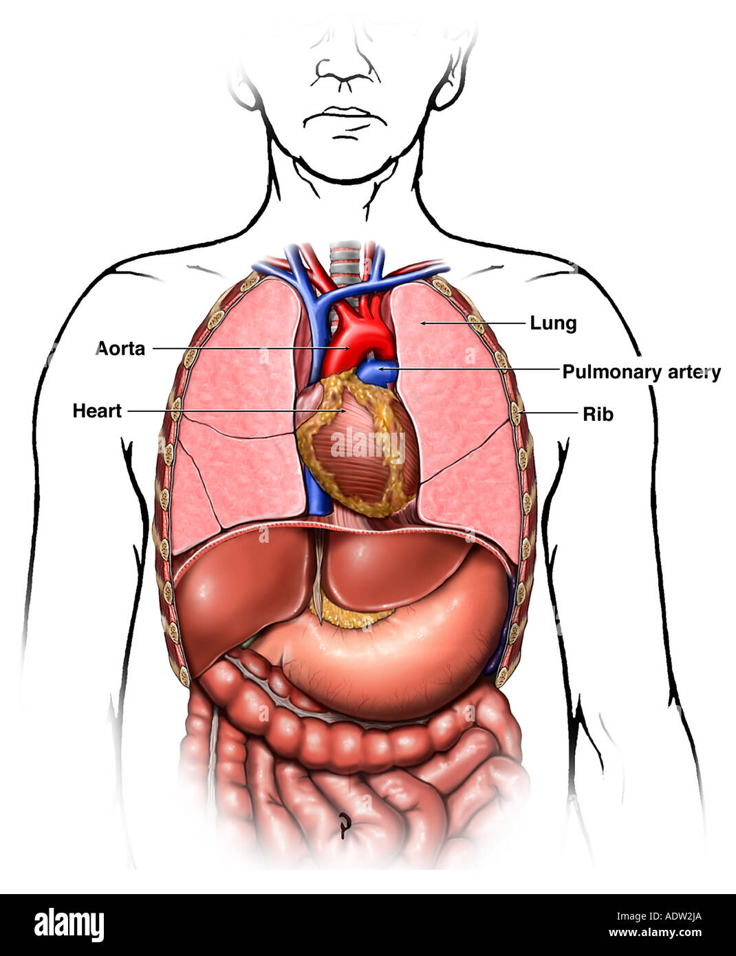
Human Chest Anatomy Images Stock Photos Vectors Shutterstock from image.shutterstock.com It is enclosed by the ribs, the vertebral column, and the sternum, or breastbone, and is separated from the abdominal cavity (the body's largest hollow space) by a muscular and membranous partition, the diaphragm. A heart attack results from blocked blood flow, often from a blood clot, to your heart muscle. See chest anatomy stock video clips. Chest a man's chest — like the rest of his body — is covered with skin that has two layers. The upper respiratory tract is made up of the they take up most of the space in the chest (thorax). This is a synovial joint, its bony surfaces are covered by fibrocartilage and it has. / upper back pain and chest pain can occur together. While it is only around one half of an inch (1 cm) in diameter, the spinal cord both carries nervous signals and processes many reflexes to support the structures of the body.
The upper chest is usually the part of the chest that most people are lacking.
It describes the theatre of events. Anatomy of the chest area. The upper posterior border of the heart is formed by the left atrium. The pectoral region is located on the anterior chest wall. Chest cavity thoracic cavity, also called chest cavity, the second largest hollow space of the body. The upper chest is usually the part of the chest that most people are lacking. Angina is the term for chest pain caused by poor blood flow to the heart. Upper back pain and chest pain can occur together. The axilla is the name given to an area that lies underneath the glenohumeral joint, at the junction of the upper limb and the thorax.it is a passageway by which neurovascular and muscular structures can enter and leave the upper limb. Anatomy of the chest and shoulder, anatomy of the chest organs, anatomy of the chest wall, anatomy of the chest wall and pleura, anatomy of upper chest area, human. I am split between the two. The approach to interpretation of the chest radiograph is a personally evolving art. Anatomy of the upper chest area :
Your torso consists of two parts — the chest and the abdomen. The upper posterior border of the heart is formed by the left atrium. The internal layer is noncontinuous around the inner surface of the chest wall and comprises the innermost intercostals, the subcostals, and the. The circulatory system does most of. Human anatomy for muscle, reproductive, and skeleton.
Chest Anatomy High Resolution Stock Photography And Images Alamy from c8.alamy.com It describes the theatre of events. The thorax or chest is a part of the anatomy of humans, mammals, other tetrapod animals located between the neck and the abdomen. The throat is one of the most complex parts of the human body. This page provides an overview of the chest muscle group. The abdomen (commonly called the belly) is the body space between the thorax (chest) and pelvis. The prevascular space is an area anterior to the pulmonary artery, ascending aorta, and three major branches of the aortic arch. Anatomy of the upper chest area. Enlargement will result in bulging of the.
Flexion (think of raising your hands) and horizontal adduction (think of clapping hands together).
Related posts of anatomy of the chest area. Chest a man's chest — like the rest of his body — is covered with skin that has two layers. It starts from the pharynx and extends to the upper end of the esophagus. Related posts of anatomy of the chest area. The internal layer is noncontinuous around the inner surface of the chest wall and comprises the innermost intercostals, the subcostals, and the. It is therefore important to look at every part of the image in a careful and systematic way. Find the perfect chest anatomy stock photo. Profile view of female chest area. The abdomen (commonly called the belly) is the body space between the thorax (chest) and pelvis. The chest contains your heart and lungs; The major muscle in the chest is the pectoralis major. The throat is one of the most complex parts of the human body. Browse 2,553 female chest anatomy stock photos and images available, or start a new search to explore more stock photos and images.
Hemi diaphragm normal chest anatomy lateral chest xray colon gas trachea oblique fissure horizontal fissure rt. Related posts of anatomy of the chest area. Anatomy of the chest area. The major muscle in the chest is the pectoralis major. The forehead is referred to as the frontal region.
Muscles Of Upper Chest And Shoulder Stock Image C020 2123 Science Photo Library from media.sciencephoto.com The pec major) is the one that commands the most real estate. The forehead is referred to as the frontal region. Anatomy of the upper chest area. This page provides an overview of the chest muscle group. Understanding the basics of throat anatomy with diagram and pictures. The epidermis is the outermost layer that provides a protective, waterproof seal over the body. Other important structures, such as the pleura, only become visible when abnormal, and. It is the part of the trunk between the neck and abdomen.
Anatomy of the chest area.
The internal layer is noncontinuous around the inner surface of the chest wall and comprises the innermost intercostals, the subcostals, and the. Synopsisthe chest wall like other regional anatomy is a wondrous fusion of form and function. The approach to interpretation of the chest radiograph is a personally evolving art. Anatomy of the chest area. Anatomy of the chest and shoulder, anatomy of the chest organs, anatomy of the chest wall, anatomy of the chest wall and pleura, anatomy of upper chest area, human. Anatomy of the upper chest area : While it is only around one half of an inch (1 cm) in diameter, the spinal cord both carries nervous signals and processes many reflexes to support the structures of the body. Anatomy of the upper chest area. Find the perfect chest anatomy stock photo. Upper can be felt in upper parts of chest, lower is in back. It is the part of the trunk between the neck and abdomen. Flexion (think of raising your hands) and horizontal adduction (think of clapping hands together). Chest wall (anterior view) therefore, the thorax can be defined as consisting of the thoracic cavity, its contents including the primary organs of the respiratory and cardiovascular systems, and the wall that surrounds it.
Share :
Post a Comment
for "Anatomy Of The Upper Chest Area / Sternum Popping Treatment Pain Chest Pain And Symptoms"


Post a Comment for "Anatomy Of The Upper Chest Area / Sternum Popping Treatment Pain Chest Pain And Symptoms"